Spinal Anatomy
The spine is made of 33 individual bones. These bones are called vertebrae. The vertebrae are supported by a number of muscles, tendons, and ligaments. There are 24 mobile vertebrae in the spine, which are separated by the inter-vertebral discs. The spine provides structural support for your body allowing movement, whilst protecting your spinal cord and the nerves.
The spine has a number of key components:
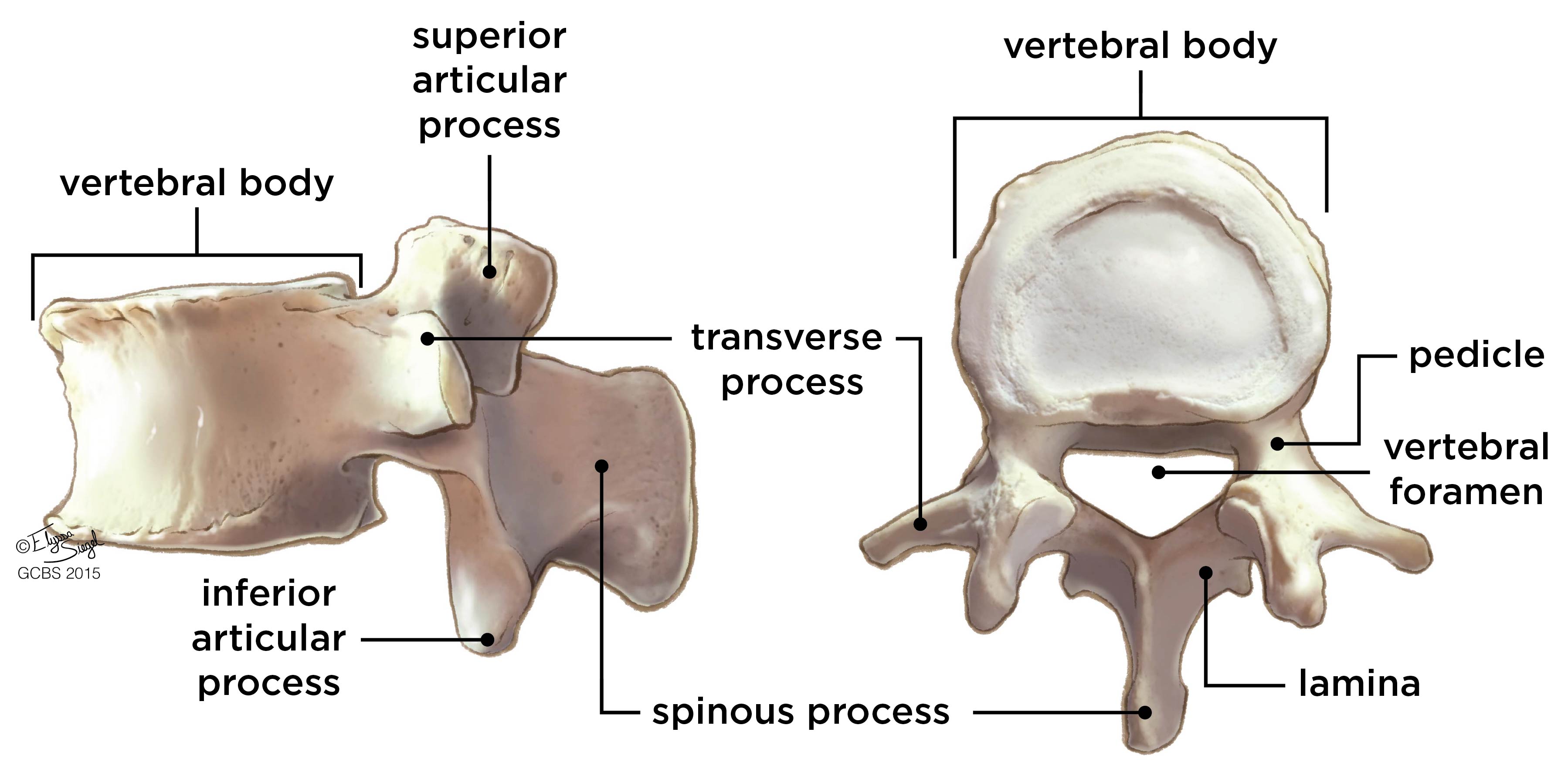
Vertebral body: The vertebral body is the front part of the vertebra. It is shaped like an ‘hour-glass’. The vertebral body and the intervertebral discs are the main weight bearing structures in the spine.
Pedicles: The pedicles are two short, rounded, bony processes that extend backwards from the vertebral body. They connect the vertebral body to the lamina. The nerves leaving the spine pass underneath the pedicles, through the exit foramen.
Lamina: The lamina is the arch of bone that protects the spinal cord at the back. There are 3 bony processes that come off the lamina: 2 articulating processes and 1 spinous process. There are 2 articulating processes, one that extends upwards and one that extends downwards. Along with the articulating processes from the adjacent levels, these articulating processes form the facet joints.
Spinous process: The spinous process is a piece of bone that comes off the back of the lamina at every level. It provides an attachment for muscles. When you feel down someone’s back, the spinous processes are the bony bits that you can feel in the middle of their spine.
Facet joints: The facet joints are formed by the articulating processes from the vertebrae above and below. These joints allow us to bend backwards and forwards as well as twist and turn. The surfaces of the facet joints are covered with cartilage. As we get older it is normal to get degenerative changes (arthritis) in the facet joints. This can cause stiffness and pain in your lower back. Arthritic facet joints can also become enlarged which can result in trapped nerves causing symptoms in your legs.

The spine seen from the side
Pars interarticularis: The pars is the part of a lamina between the superior and inferior articular processes. A stress fracture through the pars occurs in 6-8% of teenagers (the incidence is higher in very sporty teenagers). A pars fracture will usually heal with rest. A fracture or congenital anomaly of the pars in the lower lumbar spine may result in a spondylolisthesis. A spondylolisthesis is when one vertebral body slips forward relative to the vertebral body below. A spondylolisthesis can result in trapped nerves causing symptoms in your legs.
Ligamentum flavum: The ligamentum flavum is a strong ligament that connects the laminae of the vertebrae. The term “flavum” is used to describe the yellow appearance of this ligament. The ligamentum flavum stabilises the spine so that excessive motion between the vertebral bodies does not occur. As we get older, the ligamentum can become thickened. This can also contribute to nerves being trapped in your lower back causing symptoms in your legs.

Intervertebral discs: The intervertebral discs are found between each vertebral body. The discs are flat, round structures about 1cm thick. A disc is made up of a tough outer ring of tissue called the annulus fibrosis, and an inner jelly-like centre called the nucleus pulposus. The intervertebral discs separate the vertebrae, and act as shock absorbers for the spine: they compress when weight is put on them and spring back when the weight is removed. As we get older our discs become dehydrated. This is also referred to as disc degeneration. This is NOT a disease process but rather a normal part of the spine’s aging process. Disc degeneration does not directly equate to back pain.
Spinal canal: The spinal canal is where the spinal cord and the spinal nerve roots lie. The front wall of the spinal canal is formed by the vertebral body, the side walls by the pedicles and the facet joints, and the back wall by the laminae and the ligamentum flavum.
Spinal cord: The spinal cord runs within the spinal canal from the base of the brain to the level of the first lumbar vertebra. At the end of the spinal cord, the cord fibers separate into the cauda equina and continue down through the spinal canal to the sacrum.
Spinal nerves: There are thirty-one pairs of spinal nerves that branch off the spinal cord. The spinal nerves carry messages back and forth between your body and spinal cord to control sensation and movement. The spinal nerves innervate specific parts of your body. We use this pattern to diagnose the location of a spinal problem based on the distribution of pain, numbness or muscle weakness. The spinal nerves leave the spinal canal by passing through the exit foramen.
Regional Anatomy
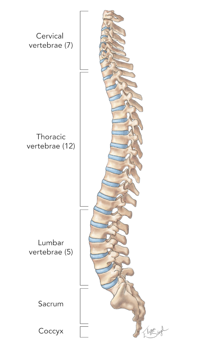
The vertebrae are named according to their location within the spine. There are 7 cervical, 12 thoracic, 5 lumbar, 5 sacral, and 4 coccygeal vertebrae. Only the top 24 bones move; the vertebrae of the sacrum and coccyx do not move. The vertebrae in each part of the spine have unique characteristic features.
The cervical spine: the main function of the cervical spine is to support the head. The 7 cervical vertebrae are labelled C1 to C7. The cervical spine is very mobile. Much of this mobility comes from the top 2 levels in the cervical spine.
The thoracic spine: the main function of the thoracic spine is to protect the organs of the chest by providing attachment for the rib cage. The 12 thoracic vertebrae are labelled T1 to T12. The thoracic spine has very limited movement.
The lumbar spine: the main function of the lumbar spine is to support the weight of the body. The five lumbar vertebrae are labelled L1 to L5. The lumbar vertebrae are the largest vertebrae in the spine, enabling them to support the weight of the body.
The sacral spine: the main function of the sacrum is to provide attachment for the pelvic bones and protect the pelvic organs. There are 5 sacral vertebrae, which are fused together.
The coccyx: the coccyx does not really have a function. There are 4 bones making up the coccyx, which are fused together. The coccyx is also known as the ‘tailbone’.
Spinal shape
In the upright position, an adult spine has an S-shape. These natural curves are maintained by the muscles. Good posture involves training your body to stand, walk, sit, and lie so that the least amount of strain is placed on supporting muscles and ligaments during movement or weight-bearing activities.
Muscles
The two main muscle groups that affect the spine are the extensor and flexor muscles. The extensor muscles enable us to stand up and lift objects. The extensors are attached to the back of the spine. The flexor muscles are in the front and include the abdominal muscles. These muscles enable us to flex or bend forward. It is important that these muscle groups are in balance with each other.
What Patients Say
Contact Us
Spire Harpenden Hospital
Amrose Lane
Harpenden
AL5 4BP
(01582) 714 304

