Spinal Investigations
In order for your back to be properly assessed you may be asked to undertake a variety of diagnostic tests. The most common tests used to diagnose back problems are X-rays and MRI scans.
X-ray
An X-ray is a painless process that uses a low dose of radiation to take a picture of your spine. X-rays show bones, but not much soft tissue. Despite high quality scans, x-rays remain useful as they provide valuable information that is not readily available from MRI or CT scans, particularly when assessing spinal deformities and abnormal movement. These x-rays are often taken with the patient standing.
If required, an x-ray can be taken at the time of your appointment.
MRI
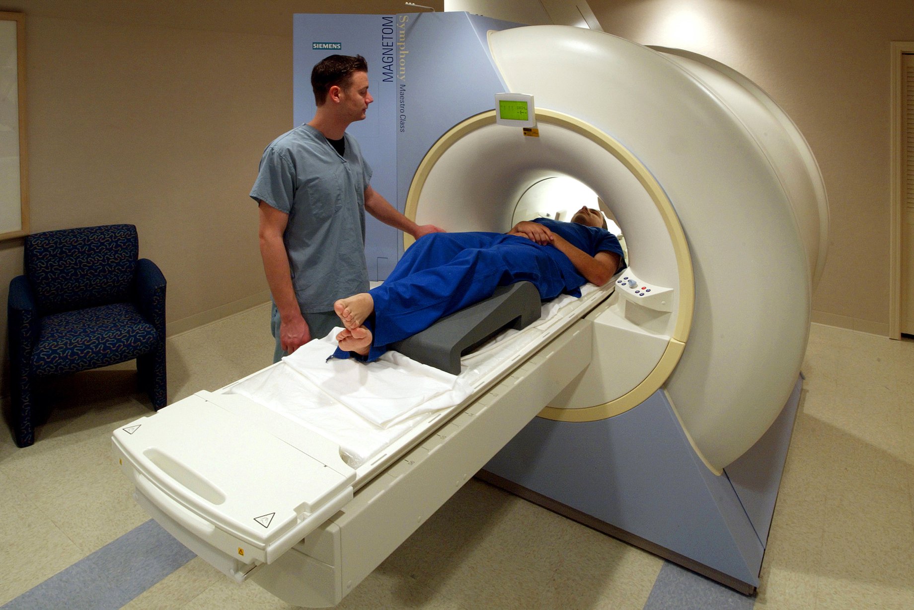
Magnetic Resonance Imaging (MRI) has revolutionized our understanding and treatment of the spine. MRI uses a powerful magnetic field and radio waves to create computer-generated images that allow us to see the spine in fine detail, enabling us to identify abnormalities of the soft tissues, such as the discs and the nerves. It is very useful for evaluating certain bony pathologies.
MRI is painless and does not involve any radiation. The only problem is the need to lie still inside, or at least partially inside, the large, cylindrical magnet where it can be a bit claustrophobic and noisy. An MRI scan normally takes about 30 minutes. It can be longer if more than one part of your spine needs to be scanned. Some patients have difficulty with having an MRI scan as they find it a very claustrophobic experience. If necessary, we can arrange for you to have your scan in an ‘open’ MRI scanner.
As a MR scanner is a large magnet, we have to make sure that there are no bits of metal in your soft tissues. We will also need to know about any recent surgery, any history of epileptic fits, if you have a pacemaker or other implanted cardiac device, of if you have hearing aids. We will ask that female patients confirm that they are not pregnant. You will be asked to fill in a pre-scanning questionnaire to ensure that there are no concerns.
You will normally be made an appointment for your MRI scan within a few days of your out-patient clinic appointment. If we are scanning one part of your body, for example your lower back, then the scan will normally take 25-30 minutes. You should expect to be at the hospital for no more than an hour. If you require more than one-part of your body to be scanned, then it will take longer. You can drive home after your scan.

CT scans
Since the advent of MRI, CT scans have been used less in the evaluation of spinal problems. As with x-rays, CT is a process that uses radiation to take a picture of your spine. The dose of radiation though is much higher than with x-rays. CT is still used to assess 3D spinal anatomy (particularly when patients have a spinal deformity or other unusual anatomy) and to evaluate certain bony pathologies.
Some patients are unable to go into a MRI scanner as they have something in their body that would be at risk if exposed to a large magnet, such as a pacemaker, cerebral aneurysm clips, or a small piece of metal lodged in the eye. In these patients CT is sometimes used as an alternative to MRI.
You will normally be made an appointment for your CT scan within a few days of your out-patient clinic appointment. The scan will normally take 10-15 minutes. You should expect to be at the hospital for no more than an hour. You can drive home after your scan.
Isotope bone scan
An isotope bone scan is a method of looking at your bones to show conditions not seen using X-rays, MRI or CT or to further evaluate something that has been seen on previous imaging, such as a potentially suspicious lesion in the bones. It requires an injection of a small amount of radioactive fluid into your arm, which is then taken up by the bones. This scan is performed three hours after the injection.
Your scan will be performed with you sitting, standing or lying down. The camera will be brought close to various parts of your body and images taken. This will take approximately 45 minutes.
There are no ill-effects from the injection. The dose of radiation received from the injection is similar to 2 years’ worth of background radiation and is not considered dangerous. However, you should avoid prolonged contact with small children and pregnant women for 24 hours following the injection. You should keep yourself well hydrated for 24-hours following you scan and empty your bladder regularly. The radioactive material will be excreted from your body in your urine. You can drive home after your scan.
SPECT CT scan
A SPECT CT scan can be a useful scan in trying to look for a source of pain, that may not be obvious on other forms of imaging. It is essentially a combination of an isotope bone scan and a special CT scan which provides more accurate information about the anatomy and function of the area being scanned and makes it easier to identify problems.
The first part of the scan – the SPECT scan – involves injecting a radioactive fluid. Once completed you can leave the department, but you will have to return 3-4 hours later when you will have a series of special CT scans which can take approximately 1 hour.
SPECT CT scanners are not available in all hospitals. I normally arrange for these scans to be done at the Royal free Hospital, Hampstead. You will get more specific information on timings when the hospital contact you to make your appointment.
There are no ill-effects from the injection. However, you should avoid prolonged contact with small children and pregnant women for 24 hours following the injection. You should keep yourself well hydrated for 24-hours following you scan and empty your bladder regularly. The radioactive material will be excreted from your body in your urine. You can drive home after your scan.
Ultrasound
An ultrasound scan is a procedure that uses high-frequency sound waves to create an image of part of the inside of the body. This image is displayed on a monitor while the scan is carried out. The scan is carried out by a Consultant Radiology doctor.
Whilst ultrasound is very commonly used in the assessment of unborn babies, it is also a valuable tool used in the assessment of musculoskeletal problems. We regularly request ultrasound scans to look at the soft tissues around the shoulder or hip, as well as in the evaluation of some lumps and swellings. If looking for shoulder or hip problems, then we may offer to carry out an injection at the time. This would be a targeted injection of a potent anti-inflammatory made up of cortisone steroid and a small amount of local anaesthetic. If we were planning for an injection, you will have been told about this beforehand.
You will normally be made an appointment for your ultrasound scan within a few days of your out-patient clinic appointment. The scan will normally take 25-30 minutes. You should expect to be at the hospital for no more than an hour. If you are having an injection then it would be sensible, though not mandatory, to bring someone with you who can drive you home afterwards.
DEXA scans
This is a scan done to assess the density (strength) of your bones. If you have reduced bone density then this is what we call osteoporosis.
When you have a DEXA scan, you will lie on your back on a flat, open X-ray table. During the scan, a large scanning arm will be passed over your body to measure bone density in your skeleton using a low dose of x-rays. The areas normally assessed are your lower back and hips. The scan usually takes 10 to 20 minutes. You can drive home after your scan.
Nerve conduction studies
A nerve conduction study is a test on the nerves in the arms and hands. It can also be done on your legs. It is a test that measures how quickly messages are travelling along your nerves. The speed at which the nerves are working can give information about whether a nerve is trapped or damaged, and where the problem may be coming from.
To obtain measurements of your nerve impulses a recording electrode will be placed onto your skin, usually on your upper and/or lower limbs. Another electrode will be used to stimulate the nerve. The stimulator produces small electrical pulses which feel like a sharp tapping sensation. The process will be repeated for a number of different nerves. Although the test can be uncomfortable, and some people experience a slight increase in their usual symptoms for a few minutes afterwards, the test will not cause you any harm.
Sometimes we also carry out a second test called EMG studies. EMG studies assess the electrical activity of the muscles. This is usually recorded using a small needle electrode inserted through the skin into the muscle, which produces a short pinprick sensation. Once in place the activity in the muscle can be observed at rest and then whilst being used.
Please advise us beforehand if you are taking any blood thinning medication or if you have a pacemaker.
The appointment will last about 45 minutes and you can drive home afterwards.
What Patients Say
Contact Us
Spire Harpenden Hospital
Amrose Lane
Harpenden
AL5 4BP
(01582) 714 304

